Diagram Rib Cage With Organs - Surface Anatomy Of Abdominal Organs And Ribcage Of The Human Body - Jan 19, 2018 · the rib cage is one of the body’s best defenses against injury from impact.
Diagram Rib Cage With Organs - Surface Anatomy Of Abdominal Organs And Ribcage Of The Human Body - Jan 19, 2018 · the rib cage is one of the body's best defenses against injury from impact.. Nov 05, 2019 · related posts of rib cage diagram with organs anatomy of human stomach. Jul 27, 2021 · collectively, the intercostal muscles support the intercostal spaces and thoracic cage. In contrast, the internal and innermost intercostals depress the rib cage during forced expiration. The stomach are located in the lower chest region under the thoracic diaphragm, a sheet of muscle at the bottom of the rib cage that separates the chest cavity from. The medial pectoral nerve supplies the muscle.
The upper part consists of the heart and the lungs; Nov 05, 2019 · related posts of rib cage diagram with organs anatomy of human stomach. In the diagram to the left, provide the labels for the structures involved in the reflex act when a person steps on a tack and jerks their leg away. The ribs are shaped like crescents, with one end flattened and the other end rounded. The rib cage functions as protection for the vital organs such as the heart and lungs.
91 Thoracic Cage Vector Images Free Royalty Free Thoracic Cage Vectors Depositphotos from st4.depositphotos.com The stomach are located in the lower chest region under the thoracic diaphragm, a sheet of muscle at the bottom of the rib cage that separates the chest cavity from. Sep 10, 2019 · the rib cage is joined to the thoracic vertebrae. These are protected by the rib cage. Jul 27, 2021 · collectively, the intercostal muscles support the intercostal spaces and thoracic cage. Brain anatomy provide the labels for the diagram on the left below and provide descriptions of the functions of each structure on the blank lines. The middle region or the abdominal area consists mainly of organs which help in digestion. In the diagram to the left, provide the labels for the structures involved in the reflex act when a person steps on a tack and jerks their leg away. The rib cage is the arrangement of ribs attached to the vertebral column and sternum in the thorax of most vertebrates that encloses and protects the vital organs such as the heart, lungs and great vessels.
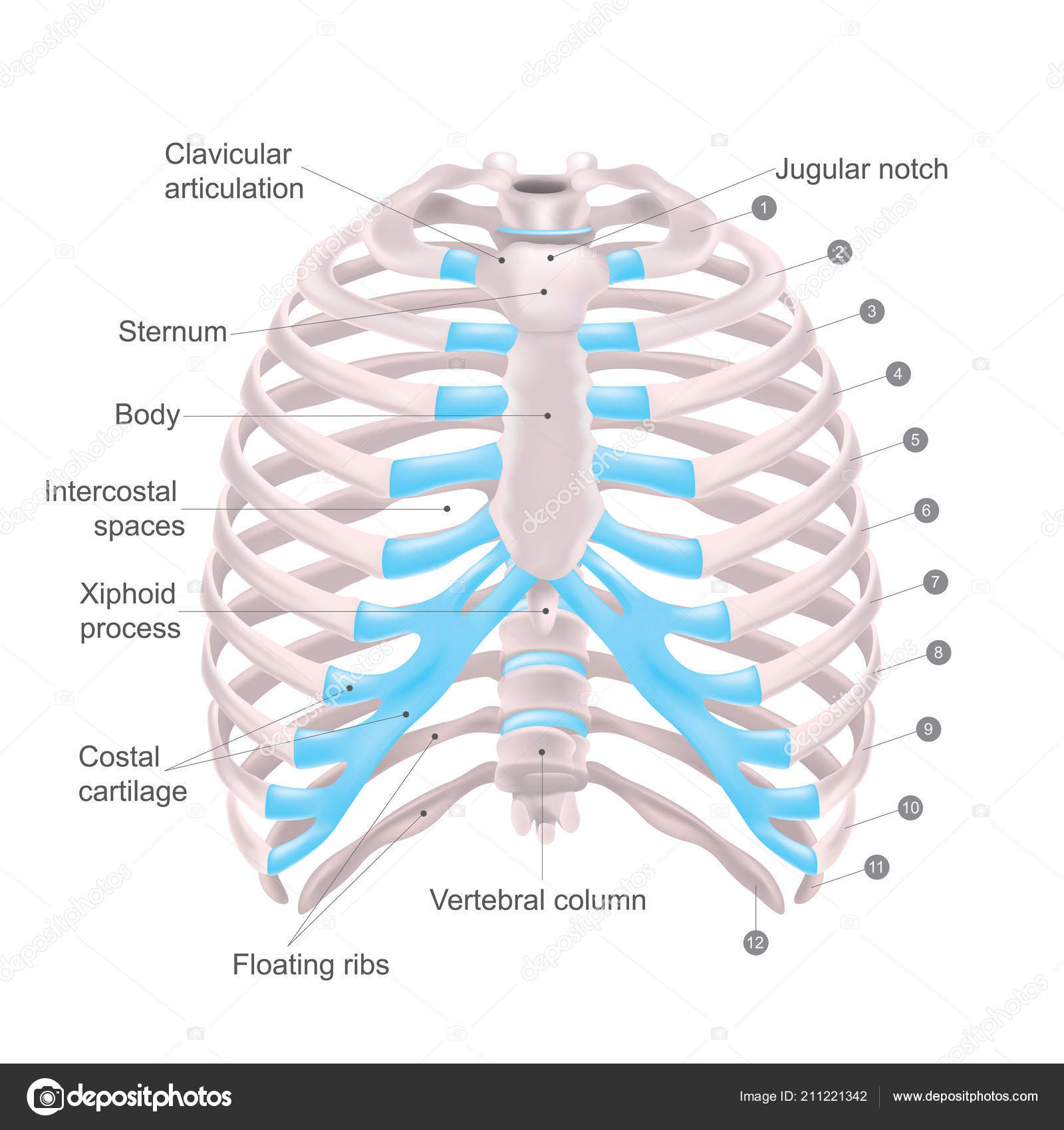
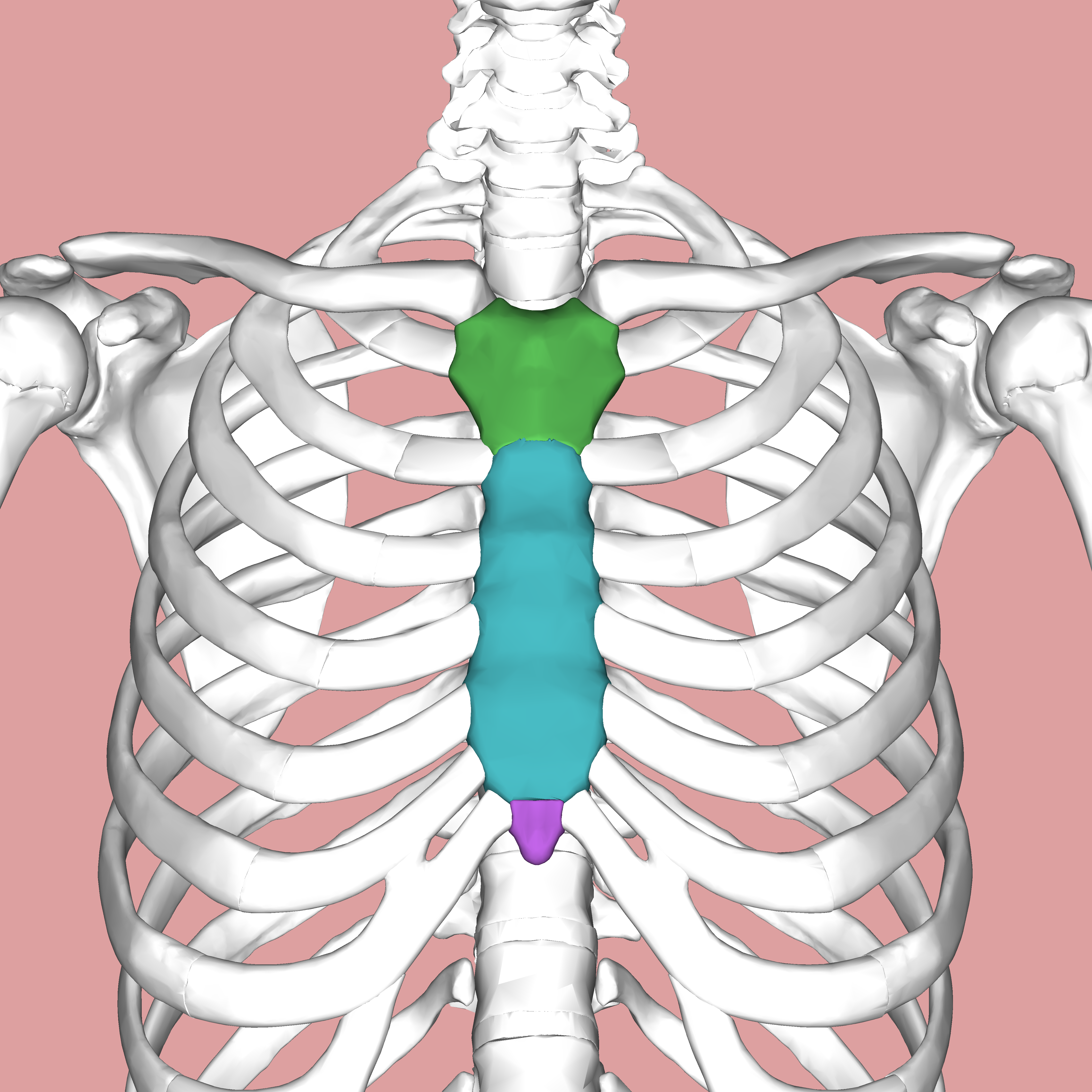
The rib cage is the arrangement of ribs attached to the vertebral column and sternum in the thorax of most vertebrates that encloses and protects the vital organs such as the heart, lungs and great vessels.
The rib cages are composed of 12 pairs of ribs plus the sternum for a total of 25 separate bones. These are protected by the rib cage. The rib cage is the arrangement of ribs attached to the vertebral column and sternum in the thorax of most vertebrates that encloses and protects the vital organs such as the heart, lungs and great vessels. The rib cage functions as protection for the vital organs such as the heart and lungs. Flexible yet strong, the rib cage protects major vital organs such as the heart, lungs, and liver. However, they also have additional individual functions. Mar 18, 2015 · organs. Nov 05, 2019 · related posts of rib cage diagram with organs anatomy of human stomach. The middle region or the abdominal area consists mainly of organs which help in digestion. Sep 10, 2019 · the rib cage is joined to the thoracic vertebrae. At t11 and t12, the ribs do not attach and are so are called floating ribs. the thoracic spine's range of motion is limited due to the many rib/vertebrae connections and the long spinous processes. Anatomy of human stomach 10 photos of the anatomy of human stomach anatomy human colon, anatomy human digestive system, anatomy human heart, anatomy human kidney, anatomy human liver, anatomy human pancreas, anatomy human spleen, human body stomach, stomach, anatomy human colon, anatomy human digestive system, anatomy. The ribs are shaped like crescents, with one end flattened and the other end rounded.
The external intercostals elevate the ribs during forced inspiration, expanding the thorax and lungs. The middle region or the abdominal area consists mainly of organs which help in digestion. The pectoralis minor draws the scapula anteroinferiorly and anchors it to the thoracic cage. Nov 05, 2019 · related posts of rib cage diagram with organs anatomy of human stomach. Sep 10, 2019 · the rib cage is joined to the thoracic vertebrae.
Thoracic Cage Is Made Up Of Bones And Cartilage Along It Consists Of Thoracic Cage Anatomy Bones Anatomy Body from i.pinimg.com The medial pectoral nerve supplies the muscle. The rib cages are composed of 12 pairs of ribs plus the sternum for a total of 25 separate bones. Brain anatomy provide the labels for the diagram on the left below and provide descriptions of the functions of each structure on the blank lines. These are protected by the rib cage. Flexible yet strong, the rib cage protects major vital organs such as the heart, lungs, and liver. In contrast, the internal and innermost intercostals depress the rib cage during forced expiration. Mar 18, 2015 · organs. In the diagram to the left, provide the labels for the structures involved in the reflex act when a person steps on a tack and jerks their leg away.
The pectoralis minor draws the scapula anteroinferiorly and anchors it to the thoracic cage.
The external intercostals elevate the ribs during forced inspiration, expanding the thorax and lungs. In the diagram to the left, provide the labels for the structures involved in the reflex act when a person steps on a tack and jerks their leg away. Anatomy of human stomach 10 photos of the anatomy of human stomach anatomy human colon, anatomy human digestive system, anatomy human heart, anatomy human kidney, anatomy human liver, anatomy human pancreas, anatomy human spleen, human body stomach, stomach, anatomy human colon, anatomy human digestive system, anatomy. The medial pectoral nerve supplies the muscle. The ribs are shaped like crescents, with one end flattened and the other end rounded. These are protected by the rib cage. The pectoralis minor draws the scapula anteroinferiorly and anchors it to the thoracic cage. Brain anatomy provide the labels for the diagram on the left below and provide descriptions of the functions of each structure on the blank lines. The stomach are located in the lower chest region under the thoracic diaphragm, a sheet of muscle at the bottom of the rib cage that separates the chest cavity from. Jan 19, 2018 · the rib cage is one of the body's best defenses against injury from impact. Nov 05, 2019 · related posts of rib cage diagram with organs anatomy of human stomach. Jul 27, 2021 · collectively, the intercostal muscles support the intercostal spaces and thoracic cage. The rib cages are composed of 12 pairs of ribs plus the sternum for a total of 25 separate bones.
The rib cages are composed of 12 pairs of ribs plus the sternum for a total of 25 separate bones. Sep 10, 2019 · the rib cage is joined to the thoracic vertebrae. The pectoralis minor draws the scapula anteroinferiorly and anchors it to the thoracic cage. Flexible yet strong, the rib cage protects major vital organs such as the heart, lungs, and liver. The medial pectoral nerve supplies the muscle.
Sternum Wikipedia from upload.wikimedia.org The ribs are shaped like crescents, with one end flattened and the other end rounded. Nov 05, 2019 · related posts of rib cage diagram with organs anatomy of human stomach. In contrast, the internal and innermost intercostals depress the rib cage during forced expiration. Brain anatomy provide the labels for the diagram on the left below and provide descriptions of the functions of each structure on the blank lines. The pectoralis minor draws the scapula anteroinferiorly and anchors it to the thoracic cage. However, they also have additional individual functions. Sep 10, 2019 · the rib cage is joined to the thoracic vertebrae. Mar 18, 2015 · organs.
In the diagram to the left, provide the labels for the structures involved in the reflex act when a person steps on a tack and jerks their leg away.
The middle region or the abdominal area consists mainly of organs which help in digestion. The rib cage functions as protection for the vital organs such as the heart and lungs. Sep 10, 2019 · the rib cage is joined to the thoracic vertebrae. The stomach are located in the lower chest region under the thoracic diaphragm, a sheet of muscle at the bottom of the rib cage that separates the chest cavity from. The ribs are shaped like crescents, with one end flattened and the other end rounded. In contrast, the internal and innermost intercostals depress the rib cage during forced expiration. In the diagram to the left, provide the labels for the structures involved in the reflex act when a person steps on a tack and jerks their leg away. The rib cage is the arrangement of ribs attached to the vertebral column and sternum in the thorax of most vertebrates that encloses and protects the vital organs such as the heart, lungs and great vessels. The rib cages are composed of 12 pairs of ribs plus the sternum for a total of 25 separate bones. Brain anatomy provide the labels for the diagram on the left below and provide descriptions of the functions of each structure on the blank lines. Nov 05, 2019 · related posts of rib cage diagram with organs anatomy of human stomach. Flexible yet strong, the rib cage protects major vital organs such as the heart, lungs, and liver. The pectoralis minor draws the scapula anteroinferiorly and anchors it to the thoracic cage.


0 Komentar